Advanced mold solution for patient-specific CRANIOPLASTY

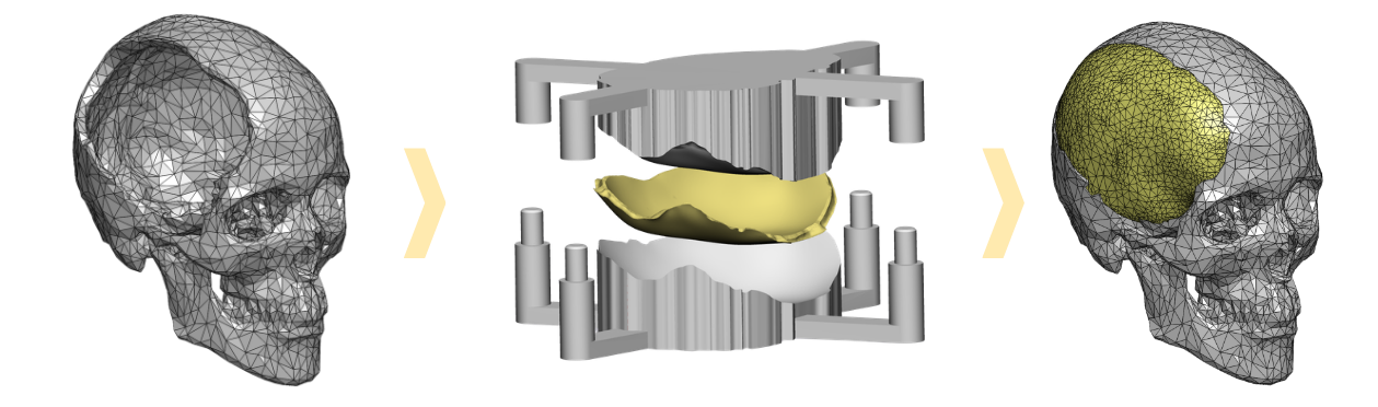
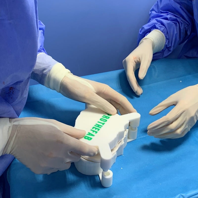
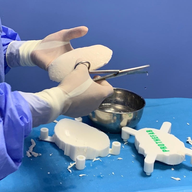
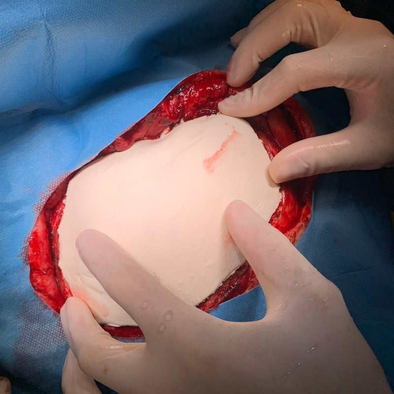
Steps of the cranioplasty mold fabrication process: 3D modeling, printed mold, implant in place

About us

Prothefab designs and manufactures patient-specific cranioplasty molds, designed for intraoperative creation of a PMMA (polymethyl methacrylate) cranioplasty implant. After a CT scan, a virtual 3D model of the patient's anatomy is generated, and the implant is precisely designed to fit the cranial defect. Using advanced 3D printing technology, Prothefab manufactures the corresponding two-part mould, which is delivered sterile to the surgical team, ready for seamless intraoperative use.

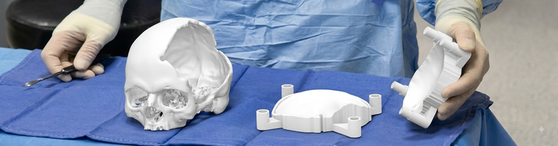
3D printed mold and skull model

How it works?

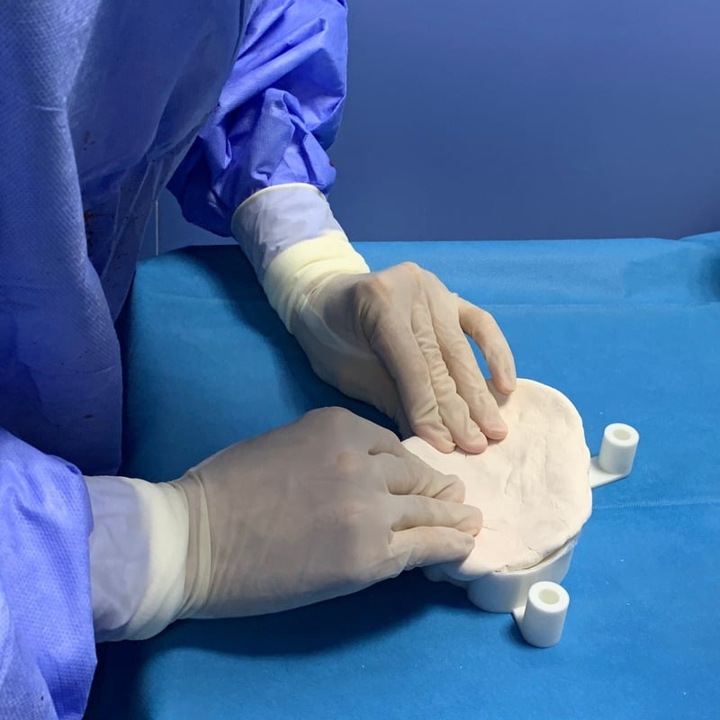
During surgery, PMMA is molded directly using the patient-specific Prothefab two-part mold, allowing precise reconstruction of the cranial defect. The implant can be easily adapted intraoperatively to accommodate anatomical variations, ensuring optimal fit, stability, and superior aesthetic results.

Why use Prothefab?

Perfect fit

Prothefab delivers highly accurate, patient-specific implants using advanced design and manufacturing technologies. Each implant restores the natural contours of the skull, providing superior aesthetic results while enhancing function. Prothefab solutions accommodate a wide range of cranial defects, from small localized areas to large, complex reconstructions.
Affordable innovation

$300 starting from

Prothefab delivers affordable, patient-specific cranioplasty solutions, ensuring high-quality reconstruction remains accessible in a wide range of healthcare environments.

Quick fabrication

48h turnaround time

Prothefab ensures rapid fabrication and shipment, enabling timely surgical planning without compromising precision or quality.

Case Studies

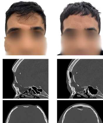
Frontal cranial defect with open sinuses

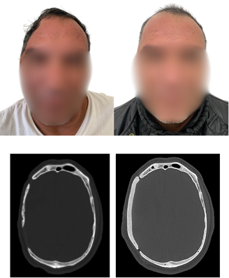
Patient before and after reconstruction, with CT scans of the frontal cranial defect

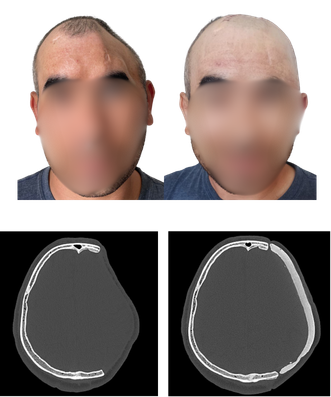
Hemicraniectomy

Patient before and after reconstruction, with axial CT scans

Hemicraniectomy

Patient before and after reconstruction, with axial CT scans

Contact us